Inotiv
CLOSE ×

Alzheimer’s Disease

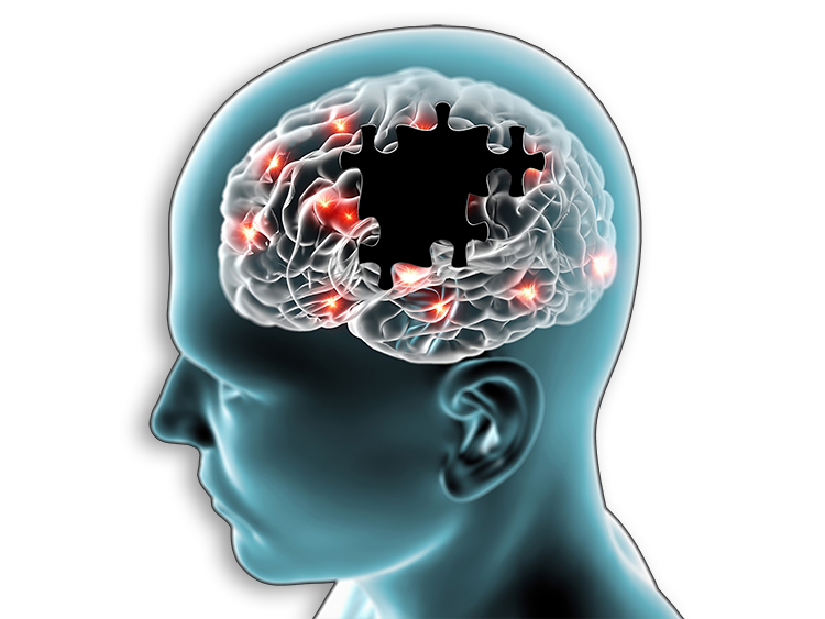
Drug development for Alzheimer’s disease has faced many obstacles. The multifaceted nature of the disease, coupled with the limited understanding of its underlying mechanisms, have presented challenges in identifying effective treatments. Additionally, clinical trials for potential drugs have had high failure rates, often due to the complexity of Alzheimer’s diagnosis and progression monitoring. These challenges highlight the urgent need for innovative approaches to better understand Alzheimer’s and develop promising therapies. Inotiv offers disease models and assays that are optimized to deliver translationally relevant data to guide your drug discovery program.

Animal Models of Alzheimer’s Disease 

Translational animal models are indispensable for understanding Alzheimer’s disease and advancing potential treatments. Alzheimer’s models offer valuable insights into the disease’s biological mechanisms, including the formation of amyloid plaques and neurofibrillary tangles, while also serving as a critical bridge between preclinical research and clinical trials to assess new therapies, as well as aiding in the exploration of reliable biomarkers for early disease detection and intervention.

Tg2576 mice

Model Summary

Tg2576 mice, acquired from Taconic Biosciences, are one of the most extensively used and well-characterized models of Alzheimer’s disease. This animal model of Alzheimer’s disease, created on a B6:SJL mixed background, carries the Swedish mutation (KM670/671NL) driven by the hamster prion protein promoter (Prnp), which causes overexpression of APP695, resulting in increased levels of amyloid-beta and formation of amyloid plaques. Tg2576 mice have been reported to exhibit cognitive deficits at 6-8 months of age, gliosis at 9 months, and amyloid plaques at 12 months. This Alzheimer’s model does not exhibit neurofibrillary tangle or neuronal loss.

Validation Data

Alzheimers-webpage-Tg2576-mice-validation-data

Tg2576 mice carrying mutations related to Alzheimer’s disease have reduced cognitive function. A) Both Alzheimer’s mutants (orange line) and normal mice (red line) learned the initial location of the hidden platform. B) The mutants (orange bars), though, did not remember the target quadrant 4 and 24 hours after the last training trial, while normal mice did (red bars). C) During reversal learning, normal mice were trained to the new quadrant (red line), but Alzheimer’s mutants failed (orange line). Data sets that do not share any letters differed significantly. (n.s.= not significant).


Alzheimers-webpage-Tg2576-mice-validation-data-2Alzheimers-webpage-Tg2576-mice-validation-data-3

Amyloid-beta was detected in perfusion fixed, paraffin embedded sections of brains (A) from female and male wildtype (WT) and Tg2576 (Tg) mice. Amyloid (brown spots) was quantified in the cortex (B) and hippocampus (C) of the mice. 

5xFAD mice

Model Summary

5XFAD transgenic mice, acquired from The Jackson Laboratory and licensed through Northwestern University, carry mutations in human amyloid precursor protein (APP) and presenilin 1 (PS1) genes associated with Familial Alzheimer's Disease (FAD). Available on both a C57BL/6J background and on a B6:SJL mixed background, the mice overexpress both mutant human APP with the Swedish (KM670/671NL), Florida (I716V), and London (V717I) FAD mutations and human PS1 harboring two FAD mutations, M146L and L286V. These mutations, driven by the mouse Thy1 promoter, contribute to an accelerated intraneuronal accumulation of amyloid-beta, amyloid deposition, gliosis, and reduced synaptic markers, along with behavioral impairments mimicking Alzheimer’s disease. This Alzheimer’s model does not exhibit neurofibrillary tangles.

ARTE10 mice

Model Summary

The ARTE10 transgenic line, created on a C57BL/6NTac background, comprises two co-integrated constructs in mice. Specifically, it involves expression of a transgene encoding the 695-amino acid isoform with the Swedish mutation of human APP (KM670/671NL), and another construct carrying human PS1 with the M146V mutation (PS1M146V). The co-inheritance of these transgenes is governed by the Thy1 promoter, allowing for streamlined breeding with other mouse models. ARTE10 mice, acquired from Taconic Biosciences, have been reported to develop amyloid plaques at 3 months of age, followed by neuronal loss, gliosis at 6 months, and cognitive impairments at 12 months of age. This Alzheimer’s model does not exhibit neurofibrillary tangles. 

Validation Data

Validation_Data_Acute_Seizure_Models

Tau, phosphorylated at residues Ser202/Thr205 (brown), was detected in the hippocampus of a 12-month-old ARTE10 mouse. The tissue was counterstained with H&E.


Validation_Data_Acute_Seizure_Models

Amyloid plaques (brown) were detected in perfusion-fixed brains from a 12-month-old ARTE10 mouse.


JNPL3 mice

Model Summary

JNPL3 mice, acquired from Taconic Biosciences, express a mutant form of human tau protein associated with neurofibrillary tangles. This mutant tau, featuring the four-repeat tau isoform (4R0N) with the P301L mutation, is regulated by the mouse Prnp promoter. The P301L mutation contributes to tau pathology and the formation of neurofibrillary tangles in these transgenic mice. JNPL3 mice develop neurofibrillary tangles at 4-6 months, and gliosis and neuronal loss at about 10 months of age. This Alzheimer’s model does not form amyloid plaques.

APPSWE-Tau mice

Model Summary

APPSWE-Tau mice, acquired from Taconic Biosciences, are produced through the mating of Tg2576 mice, which carry the Swedish mutation (KM670/671NL) of human APP, and JNPL3 mice, which express the four-repeat tau isoform (4R0N) with the P301L mutation. These mice exhibit neurofibrillary tangles and gliosis at about 3 months of age, and amyloid plaques at 9 months. There are no reports of APPSWE-Tau mice displaying synaptic loss or behavioral impairments.

Tau (P301S) PS19 mice

Model Summary

Tau P301S (PS19) mice from The Jackson Laboratory express a human tau protein with the disease associated P301S mutation and that is under the control of the mouse Prnp promoter. The mice exhibit gliosis at 3 months of age, followed by synaptic loss at 3-6 months and neurofibrillary tangles and cognitive impairments at about 6 months. There are no reports of Tau P301S (PS19) mice displaying amyloid plaques.

Genetically Engineered Alzheimer's models

Our pre-developed transgenic rat models express genetic mutations associated with Alzheimer’s disease and provide a valuable platform to study Alzheimer’s disease mechanisms, test therapeutic interventions, and bridge the translational gap between preclinical findings and potential human treatments.


Not seeing the model you need? Contact us to discuss developing a new disease pharmacology model for your preclinical drug discovery program.

Behavioral Tests for Alzheimer’s Models

Behavioral assays are crucial in the preclinical testing of new therapies for Alzheimer’s disease as they help establish the validity of disease models, screen potential drug candidates, optimize dosing, understand mechanisms of action, and provide insights into the long-term effects of therapies. We offer a comprehensive suite of validated assays to assess the effects of potential treatments on the cognitive and behavioral functions of Alzheimer’s models.

  • Open field assay
  • Gait analysis
  • Elevated plus maze
  • Barnes maze
  • Morris water maze
  • Contextual fear conditioning


Download our webinar, Alzheimer's and Parkinson’s Disease In Vivo Models for Drug Development, to hear Inotiv scientists compare the various models available for AD and PD drug development and the behavioral, molecular, and histopathological endpoints that can be used to determine if treatments show enough therapeutic potential to progress through the pharmaceutical development pipeline.

Featured speakers include Inotiv's industry experts Lauren E. Hood, PhD (left) and Clark W. Bird, PhD, MBA (right).

Lauren-Clark-ALZ-Webinar-presenters


Brain Histopathology Services

Histopathology of animal models of Alzheimer’s disease is vital for therapy development, confirming model relevance, tracking disease progression, and identifying treatment targets. Insights into mechanisms of action gained from animal models, as well as the assessment of treatment efficacy, dosage optimization, and biomarker discovery, streamline the translation of research findings to clinical trials, bridging the gap between preclinical investigation and human interventions. We offer a full range of immunohistochemistry, histopathology, and image analysis/digital pathology services to evaluate pathological features of this disease in your Alzheimer’s animal model, including plaque formation, microglial activation, and synaptic loss.

  • Amyloid-beta plaques
  • Microglial activation
  • Astrocyte activation
  • Synaptic loss

Additional Endpoints for Alzheimer’s Disease Research

Our capabilities extend beyond models and behavioral testing. We have a broad range of GLP and non-GLP in vivo and in vitro assays that can be customized to provide solutions for your drug discovery program.

  • Stereotaxic surgery
  • Tissue harvesting
  • Oxidative stress enzymology
  • Neurotransmitter and metabolite quantification

Subscribe to updates from Inotiv

Sign up and you’ll receive the latest updates that matter to you, delivered straight to your inbox.

Subscribe now  
Alzheimer's Disease