Parkinson’s Disease
Translational In Vivo Models and Assays for Drug Discovery

Parkinson’s disease is a progressive nervous system disorder that causes uncontrollable movements due to degradation of midbrain dopaminergic neurons. It is a complex condition thought to be caused by multiple factors including genes, toxins, and environmental factors, though its exact etiology is still unknown. Treatments currently available for Parkinson’s disease only ease symptoms; new therapeutics that slow or stop disease progression are still needed. Our portfolio of translational in vivo models for Parkinson’s disease and team of scientists experienced at assessing behavioral, biochemical, and histological endpoints, can support your drug discovery studies.
Animal Models of Parkinson’s Disease
We offer multiple animal models of Parkinson’s disease that simulate the behavior and monoamine depletion seen in the human disease. Our induced Parkinson’s models can be generated in both rats and mice.
6-OHDA-induced Parkinson’s model
Model Summary
For the 6-OHDA-induced Parkinson’s model, rodents receive a unilateral injection of 6-hydroxydopamine (6-OHDA) into the medial forebrain bundle (MFB) to selectively destroy the catecholaminergic neurons of the nigrostriatal pathway. These animals exhibit motor impairments that mirror Parkinson’s disease symptoms.
Study Design

Validation Data

Expression of tyrosine hydroxylase (TH) was detected in a perfusion fixed, paraffin embedded section of brain from a rat that received a unilateral injection of 6-OHDA into the MFB on the right side. TH-positive neurons (brown staining) were only detected in the side contralateral to the lesion.

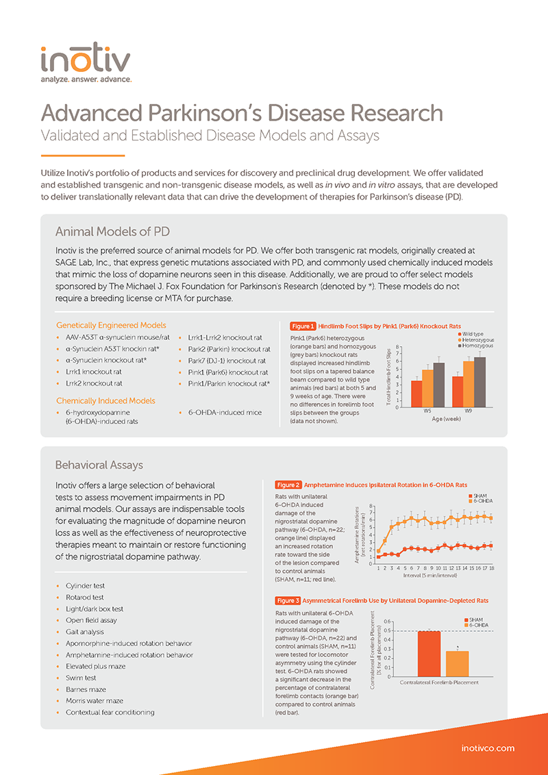
Rats with unilateral 6-OHDA-induced lesions of the nigrostriatal dopamine pathway showed a significant decrease in the percentage of contralateral forelimb contacts (orange bar) compared to control animals (red bar) in a cylinder test.
AAV-A53T α-synuclein Parkinson’s model
Model Summary
For the AAV-A53T α-synuclein Parkinson’s model, rodents overexpress a mutated form of α-synuclein (A53T) in midbrain dopaminergic neurons following delivery of an adeno-associated virus (AAV)-A53T α-synuclein vector into the substantia nigra (SN).
Study Design

Validation Data

Expression of phospho-α-synuclein was detected in a perfusion fixed, paraffin embedded section of brain from a Parkinson’s mouse model. The Parkinson’s mouse model received a unilateral injection of AAV-A53T α-synuclein-A53T. Phospho-α-synuclein was detected in the substantia nigra on the ipsilateral side of the AAV-A53T α-synuclein injection.
Genetically engineered Parkinson’s models
Models
Our predeveloped transgenic rat models express genetic mutations associated with Parkinson’s disease.
Download our webinar, Alzheimer's and Parkinson’s Disease In Vivo Models for Drug Development, to hear Inotiv scientists compare the various models available for AD and PD drug development and the behavioral, molecular, and histopathological endpoints that can be used to determine if treatments show enough therapeutic potential to progress through the pharmaceutical development pipeline.
Featured speakers include Inotiv's industry experts Lauren E. Hood, PhD (left) and Clark W. Bird, PhD, MBA (right).

Behavioral Tests for Animal Models of Parkinson’s Disease
We provide behavioral tests for assessing movement impairments in Parkinson’s models and the effectiveness of neuroprotective therapies.
- Cylinder test
- Rotarod test
- Light/dark box test
- Open field assay
- Gait analysis
- Apomorphine-induced rotation behavior
- Amphetamine-induced rotation behavior
- Elevated plus maze
- Swim test
- Barnes maze
- Morris water maze
- Contextual fear conditioning
- Histopathological assessment
- Stereotaxic surgery
- Tissue harvesting
- Oxidative stress enzymology
- Neurotransmitter and metabolite quantification
- CBC/clinical chemistry analysis
- Mass spectrometry proteomics
- Primary neural cell culturing
- Human stem cell and brain organoid culturing
- Confocal and electron microscopy
Not seeing the model you need? Contact us to discuss developing a new disease pharmacology model for your preclinical drug discovery program.


