Diabetic and Metabolic research solutions
Inotiv
CLOSE ×

Osteoarthritis

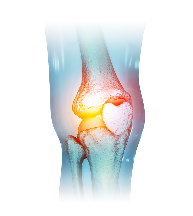
Drug discovery for osteoarthritis (OA) is crucial due to the significant impact of the disease on joint function, mobility, pain, and quality of life, affecting a large portion of the population. Currently, there are limited effective pharmacological interventions, emphasizing the urgent need for novel therapeutic options to alleviate symptoms and modify the course of the disease. Advancements in drug discovery not only address the unmet medical needs in managing OA but also contribute to reducing the socioeconomic burden associated with this prevalent musculoskeletal condition.

In Vivo OA Models

OA models, whether naturally occurring or induced through various methods, provide diverse avenues for studying cartilage degeneration and potential therapies. While spontaneous OA is species-specific, induced OA can be generated in many species. Surgically induced OA models share similarities with human or animal disease. Our scientists are experts in modeling OA joint damage and pain using a variety of induction techniques, giving you a range of options for choosing the best OA model to assess your novel therapeutic.

Medial Meniscal Tear OA Model

Model Summary

The medial meniscal tear (MMT) OA model involves cutting the medial meniscus so that the meniscus no longer protects the medial surfaces of the tibial plateau and femoral condyle, mimicking a common injury seen in humans. Mice, rats, and guinea pigs are used to create an MMT OA model, with rats being the most utilized species. However, a benefit to using guinea pigs for the MMT OA model is that they will develop spontaneous OA. Thus, the contralateral non-operated joint in a guinea pig MMT OA model can be utilized in the evaluation of effects of various treatments.

The induced meniscal tear in the MMT OA model leads to altered biomechanics and increased stress on the articular cartilage within the joint causing progressive cartilage degeneration. By 3- to 6-weeks post-surgery, tibial cartilage degeneration may be focally severe on the outer one-third of the tibia (and less severe in the middle and inner areas). Additionally, osteophytes form and progressively increase in size, and substantial subchondral bone changes occur in the medial tibia beneath the cartilage lesions, allowing assessment of both chondroprotective and bone effects. Of the major microsurgery models, the MMT OA model has been shown to be a more reliable model than the anterior cruciate ligament (ACL) transection-induced arthritis model, which induces excessively severe lesions with much greater variability.

Validation Data

MMT_stained-sections_28days-63days


Representative photomicrographs of knee joint sections stained with toluidine blue from rat MMT OA models (right column) 28-days (top row) and 63-days (bottom row) post-surgery. Stained sections of knee joints from age-matched, normal rats (left column) shown for comparison to illustrate disease progression.


Osteoarthritis-photomicrographs-of-knee-joint-sections


Representative photomicrographs of knee joint sections, stained with toluidine blue, from a normal rat (A) and a rat MMT OA model (B; 28-days post-surgery), depicting the division of the tibial plateau into 3 zones of equal width using an ocular micrometer, with zone 1 on the outside and zone 3 on the inside. (C) Section of a knee joint from a rat MMT OA model, stained with toluidine blue, depicting substantial cartilage degeneration scoring.

Related Articles


Destabilization Of The Medial Meniscus OA Model

Model Summary

Destabilization of the medial meniscus (DMM) OA model involves inducing structural changes in the knee joint by surgically cutting the medial meniscotibial ligament, leading to destabilization of the medial meniscus. Conducted in either mice or rats, this manipulation induces mild to moderate OA lesions in the central weight bearing area of the medial femoral condyle and medial tibial plateau. Lesions are evident as early as 2-weeks post-surgery and increase in severity over time at a slower rate than in the MMT model. Of the major microsurgery models, the DMM OA model has been shown to be a more reliable model than the ACL transection-induced arthritis model, which induces excessively severe lesions with much greater variability.

Validation Data

Osteoarthritis-photomicrographs-of-knee-joint-sections-2

Representative photomicrographs of knee joint sections stained with toluidine blue from a rat MMT OA model 28-days post-surgery (A) and from a rat DMM OA model 56-days post-surgery (B).


DMM_bar-graph

Mean cartilage degeneration scores for three equally spaced regions across the medial tibial plateau surface, Zone 1 (outside region, red bars), Zone 2 (middle region, orange bars), Zone 3 (inside region, light orange bars), along with the sum of the three areas (grey bars), for a rat MMT OA model 28-days post-surgery (A) and rat DMM OA model 56-days post-surgery (B).

Monoiodoacetate-Induced OA Model

Model Summary

The monoiodoacetate (MIA)-induced OA model involves injecting MIA, an inhibitor of glycolysis, into a joint, typically the knee, to kill chondrocytes, leading to an acellular matrix and eventual collapse of the cartilage matrix into the epiphysis. This model is biphasic as it simulates early joint pain due to inflammation, and then severe cartilage degradation and bone changes, which is seen in humans with end-stage OA. This model has diverse applications. Researchers employ this model to study the pathogenesis of OA pain and gain insights into the underlying mechanisms of joint degeneration. Additionally, this model can be used to assess agents designed to hinder acute proteoglycan loss via aggrecanase or MMPs, impede collagen matrix degeneration through collagenase, and induce repair. It is also valuable for evaluating the impact of agents on bone remodeling given its propensity to result in sclerotic subchondral bone lesions and osteophyte formation. Additionally, there is substantial arthrofibrosis, making this model a good tool for evaluating methods to inhibit this process.

Validation Data

Osteoarthritis-rat-knee-joints

Rat knee joints, stained with toluidine blue, from rats injected intra-articularly with saline (A), 0.5 mg of MIA (B) or 2.0 mg of MIA (C). The lower dose of MIA results in milder, more variable lesions whereas the higher dose of MIA results in severe, more consistent lesions.

MIA_line-graph

Weight bearing asymmetry, a translatable measure of joint pain, was assessed in a rat model of MIA-induced OA (red boxes) and control animals (grey boxes). Rats treated with MIA exhibited differences in weight exerted by its hind paws, with the untreated (left) limb wielding more weight than the treated (right) limb. There was little difference in weight exerted by the hind paws of the control rats.

Peptidoglycan Polysaccharide-Induced OA Model

Model Summary

The peptidoglycan polysaccharide (PGPS)-induced mono-articular pain model involves injecting PGPS, a component of bacterial cells walls, into a joint space, which triggers an inflammatory response. The phenotype can then be flared 2-weeks to months later with an IV injection of PGPS. This model provides a robust way to look at intra-articular therapies and their effects on inflammatory pain, similar to what is seen in an OA flare. OA is primarily considered a degenerative joint disease, but inflammation is believed to play a role in pain pathogenesis by releasing inflammatory mediators, such as cytokines and prostaglandins, which lead to pain. Synovial inflammation and alterations in subchondral bone density can further contribute to joint damage, and inflammation can sensitize nerve endings, increasing pain perception and potentially influencing joint use patterns.

Partial Medial Meniscectomy-Induced OA Model

Model Summary

The partial medial meniscectomy-induced OA model is used to investigate the mechanisms and development of OA following partial medial meniscectomy, providing valuable insights for research purposes. It involves surgically removing approximately half of the anterior portion of the medial meniscus in animal subjects, often mature Beagle dogs. This controlled injury aims to mimic conditions leading to degenerative changes in the knee joint, including cartilage degradation. The surgical procedure allows researchers to study the progression of OA over a specific timeframe. Despite the animal's attempts at meniscus regeneration, induced lesions consistently exhibit moderate degenerative changes in the tibial and femoral cartilage.

Partial Lateral Meniscectomy-Induced OA Model

Model Summary

The partial lateral meniscectomy-induced OA model is an experimental method used to study OA in animal subjects, often in rabbits as they preferentially load the lateral aspect of the knee joint. In this model, a surgical procedure is performed to partially remove the lateral meniscus, a cartilaginous structure in the knee joint. The surgery induces a consistent focal degenerative change that involves about one-half of the lateral tibial plateau and femoral condyle, mimicking conditions that may lead to OA. This model provides a controlled environment for researchers to explore the mechanisms and development of OA following partial lateral meniscectomy and test potential chondroprotective agents.

Related Articles

Canine Anterior Cruciate Ligament Transection-Induced OA Model

Model Summary

Transection of the anterior cruciate ligament (ACL) results in a true instability-induced OA lesion, mimicking natural OA in canines and humans after traumatic injury. Clinically these lesions progress to OA in both species after extended periods, providing an opportunity to study slowly progressing OA. Large hounds, rather than Beagle dogs, must be used to reliably generate typical OA lesions in a reasonable period of time. Animals tend to favor the limb post-surgery, perceiving instability, and compensating with reduced load bearing. While they may develop OA lesions over time, early changes are mild and variable, limiting efficiency in compound testing.

Spontaneous OA Model

Model Summary

Spontaneous OA manifests in the medial compartment of the knee joint in a variety of guinea pig strains. While occurring in both genders, males exhibit faster growth, reaching higher body weights and displaying more consistent pathological alterations. Typically, the disease is bilaterally symmetrical in terms of incidence and severity. Initial changes, observed around 3-months of age (700 grams weight), involve focal chondrocyte death, proteoglycan loss, and fibrillation in the medial tibial plateau. OA pathogenesis progresses, and by 1-year, profound cartilage degeneration, subchondral sclerosis, bone cysts, and severe meniscal degeneration has occurred. The predictability and similarity to human disease make this guinea pig model valuable for studying OA pathogenesis and potential therapeutic interventions.

Validation Data

Spontaneous-OA_images

Knee joint from 4-month-old guinea pig (A). Photomicrographs of toluidine blue-stained sections of the medial aspect of a knee joint obtained from a 6-month (B), 9.5-month (C) and 18-month-old (D) guinea pig. Images illustrate progression of cartilage degeneration on the tibial plateau.

Related Articles

Download our webinar, Exploring Animal Models for Osteoarthritis Drug Development, where our scientists examine a range of animal models used in OA drug research and compare the lesion morphologies and pathological mediators present in these models.

Featured speakers include Inotiv's industry experts Jed Pheneger, Senior Director of Pharmacology — Inflammation and Allison Bendele, DVM, PhD, DACVP Pathologist

exploring-animal-Models-for-Osteoarthritis-Drug-Development-speakers


Assays for Evaluating OA-Induced Pain and Inflammation

Behavioral assays are commonly employed for evaluating pain and inflammation in OA models. These assessments play a crucial role in the development of new drugs for OA by providing insights into the effectiveness of potential therapies in alleviating pain and improving joint function.

  • Bone pain assessment
  • Gait analysis
  • Inflammatory pain assessment
  • Knee swelling
  • Von Frey analysis
  • Weight bearing analysis


Not seeing the model you need? Contact us to discuss developing a new OA model for your OA drug development program.

Histopathological Analysis in OA Models

Histopathological analysis in OA models is vital for understanding structural changes in joint tissues. Beyond revealing the severity and progression of OA, this analysis enhances study reliability by validating and correlating findings from other assays. It is also crucial for evaluating the efficacy of potential therapeutic interventions, offering insights into treatments' impact on joint damage mitigation, inflammation reduction, and tissue repair promotion. Learn more on how tissue from MMT, MIA, and DMM OA models are processed and analyzed by a board certified veterinary pathologist.

Additional Endpoints

Our capabilities include a broad range of GLP and non-GLP in vivo and in vitro assays that can be customized for your needs. The combination of these diverse assays provides a comprehensive and multifaceted approach to understanding the complex interplay of pain and inflammation in OA, facilitating the evaluation of potential therapeutic interventions.

  • PK/PD blood collections
  • Cytokine/chemokine analysis with ELISA and Luminex® assays
  • CBC/Clinical chemistry analysis
  • Soft tissue collection
  • Immunohistochemistry analysis

Frequently Asked Questions (FAQs) for OA Drug Discovery

Why Use Rats To Develop OA Models

One advantage of using rats to develop an OA model is that results can be obtained in relatively shorter periods and the response of the animals to surgery is very consistent. Additionally, rat models are commonly used in toxicology testing. Efficacy in this species in combination with toxicology evaluation allows for the generation of a therapeutic index for compounds under evaluation.

Related Articles

Subscribe to updates from Inotiv

Sign up and you’ll receive the latest updates that matter to you, delivered straight to your inbox.

Subscribe now